Ultrasound Adds Real Time Information
Seeing Beneath the Surface
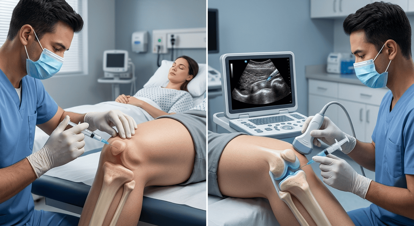
Traditional injectable treatment relies on facial examination, anatomy knowledge, palpation, and technique. With ultrasound-guided injectables, the practitioner can also view structures beneath the skin in real time. The image may help identify vessels, tissue layers, and existing filler deposits. This additional information can support planning in selected facial areas. However, ultrasound does not replace clinical judgement or anatomical expertise. It becomes another tool within a complete safety process.
High frequency ultrasound produces an image through sound waves rather than radiation. The practitioner moves a probe across gel placed on the skin. Doppler settings may help show blood flow within visible vessels. Image quality depends on equipment, training, and correct interpretation. A poorly understood scan can create false confidence. Therefore, ultrasound use should be paired with substantial hands on education.
Pre-Treatment Mapping Can Change the Plan
Anatomy Is Not Identical in Every Face
Textbook vessel patterns provide guidance, but individual anatomy can vary. Before ultrasound-guided injectables, scanning may reveal a vessel in an unexpected position. The provider can then alter the entry point, depth, or product placement. They may also decide that a proposed treatment area carries unnecessary risk. This information is particularly valuable in complex or previously treated faces. A safer plan sometimes means performing less treatment.
Traditional injectors also reduce risk through careful technique and conservative volumes. They assess movement, skin thickness, landmarks, and previous treatment history. Many routine injections can be performed safely without live imaging. Ultrasound is not mandatory for every low risk or superficial procedure. Its value rises when anatomy is uncertain or precision is especially important. The choice should be based on clinical need rather than novelty.
Placement Can Be Confirmed More Precisely
Depth and Product Location Matter
Injectables behave differently when placed in different tissue planes. During ultrasound-guided injectables, the provider may follow the needle or cannula and observe product placement. This can help confirm that treatment reaches the intended layer. It may be useful near vessels, glands, muscles, or delicate structures. Precise placement can support a more predictable correction with conservative product use. Still, the final result also depends on product choice and artistic judgement.
Traditional techniques often use reliable landmarks and tactile feedback to select depth. Experienced injectors can achieve excellent results through these established methods. The limitation is that they cannot directly see every hidden structure. Ultrasound provides information, but it can slow the appointment and increase cost. Some areas are also technically difficult to scan and inject simultaneously. Patients should understand both the advantages and practical limitations.
Previous Filler Can Be Easier to Investigate
Old Product May Not Be Where Expected
Clients do not always remember which filler was used or where it was placed. With ultrasound-guided injectables, a practitioner may identify deposits, migration, or areas of product accumulation. This information can prevent adding filler into an already crowded space. It may also guide targeted hyaluronidase when suitable hyaluronic acid filler needs dissolving. Not every material appears the same on ultrasound. Interpretation should therefore remain cautious and linked to clinical findings.
Traditional assessment may detect visible lumps or irregularity through touch and examination. However, deeper product can be difficult to locate confidently. Blind dissolving may expose normal tissue to unnecessary enzyme. Imaging can help make the approach more selective in complicated cases. Even then, dissolving carries risks and may need more than one visit. A detailed treatment history remains valuable whenever it is available.
Ultrasound Supports Safety but Cannot Remove Risk
Training Still Matters Most
Vascular occlusion is a rare but serious complication of dermal filler treatment. Careful ultrasound-guided injectables may help practitioners map vessels and investigate suspected problems more accurately. The technology may also support rapid, targeted management when complications occur. Yet no scan can guarantee that an injection will be risk free. The provider must still recognise symptoms and act without delay. Emergency supplies and a clear complication protocol remain essential.
Patients should ask who performs the scan and how often they use ultrasound. The practitioner should explain their medical qualifications and emergency training. They should also discuss product type, consent, and possible adverse effects. A clinic should never use imaging as a substitute for honest risk communication. Confidence should come from systems, skill, and preparedness together. That combination is more reassuring than technology alone.
The Best Method Depends on the Situation
Choose Precision Where It Adds Value
Simple treatments in familiar areas may not require live imaging. However, ultrasound-guided injectables can be particularly useful for high risk zones, uncertain anatomy, previous filler, or complication management. Some practitioners use ultrasound only for mapping before injection. Others use it dynamically throughout selected parts of the procedure. Both approaches can be reasonable when supported by training. The patient should understand how imaging changes the proposed plan.
A thoughtful consultation should compare benefits, cost, time, and alternatives. Ask why ultrasound is recommended for your specific concern. The answer should identify a practical safety or precision advantage. If imaging is unavailable, ask how the clinic manages anatomical risk. Traditional and image guided techniques can both produce excellent results. The safest choice remains a qualified practitioner who treats conservatively and communicates clearly.
Not sure what your skin needs?
Book a consultation at our Clifton clinic and our doctors will build a plan around your skin.